 |
CANCER BIOPHYSICS LAB |
“Biomedical Engineering can improve human lives”Dr. Triantafyllos Stylianopoulos is an Associate Professor and Head of the Cancer Biophysics Laboratory at the University of Cyprus. He was trained as a Chemical Engineer at NTUA, Greece (Diploma, 2003) and at the University of Minnesota, USA (PhD, 2008) and did his postdoctoral training at the Massachusetts General Hospital and Harvard Medical School in Boston, USA (2008-2010). Dr. Stylianopoulos has published >150 peer-reviewed scientific articles (h-index: 59, >20,500 citations) in the fields of cancer research, nano-immunotherapy and bioengineering/biomechanics. His research team has developed novel therapeutic strategies to remodel the tumor microenvironment in order to improve the delivery and efficacy of cancer therapeutics. These strategies have already made it to the clinic, through initiated clinical trials. In recognition of his work, Dr. Stylianopoulos has been awarded the Y.C. Fung Early Career award by the Bioengineering Division of the American Society of Mechanical Engineers (2016), as well as the Most Cited Paper Award (2014) and the Best Paper Award (2019) by the USA Biomedical Engineering Society. Dr. Stylianopoulos has been also the recipient of the UNESCO-Equatorial Guinea International Prize for Research in the Life Sciences (2024) as well as the Young Researcher (2014) and Distinguished Researcher Award (2022) by the Cyprus Research and Innovation Foundation. The Cancer Biophysics Lab has secured >€10M in research funding the last 10 years from the European Commission, the National Institutes of Health (USA) and the European Research Council-ERC. These grants include the highly selective ERC Starting, ERC Consolidator and ERC Advanced grants and 3 ERC Proof of Concept grants. His efforts towards research commercialization have resulted in 3 recent patents, a license agreement with Materia Therapeutics (NV, USA) for transfer of Intellectual Properties (2023), a research agreement with NanoCarrier Ltd (Japan) and a collaboration with Neobe Therapeutics (UK) to evaluate their drug formulations.
Professor Triantafyllos Stylianopoulos Cancer Biophysics Lab Department of Mechanical and Manufacturing Engineering, University of Cyprus
Cancer MechanobiologyAs tumours grow, they generate mechanical forces due to increased cell proliferation and extracellular matrix remodelling. This results in increased pressure within the tumour and blood vessel constriction, impairing perfusion and drug delivery. Losartan, an anti-hypertensive drug, can alleviate mechanical forces and improve chemotherapy response in cancer patients. However, the remaining mechanical forces induce chemotherapy resistance, which we refer to as mechanoresistance. We aim to address chemotherapy resistance caused by these mechanical forces. We employ preclinical tumor models to investigate the underlying mechanisms using cutting-edge bioengineering and biology techniques. Following validation of these findings in human cells, novel inhibitors will be tested in combination with losartan and immunotherapy as an improved anti-cancer strategy. Specifically, we develop multicellular tumor spheroids (MCS) grown within an agarose gel to create physiologically relevant systems of mechanical compression of tumors. In parallel, human samples from patients are collected. The stiffness (i.e., elastic modulus) of the agarose is measured with mechanical testing, whereas the stiffness of murine and human tumors are measured with ultrasound shear wave elastography prior to tumor removal. With the use of computational modelling, stiffness data are converted |
The Cancer Biophysics Laboratory is a vibrant and internationally competitive research environment where novel computational approaches are combined with state-of-the-art experimental techniques to further explore the mechanopathology of cancer and overcome the barriers to the effective delivery of drugs to solid tumours. The focus of the lab is the application of principles from engineering and biology in order to investigate the mechanisms with which physical forces are related to tumour growth, progression and treatment. At the Cancer Biophysics Laboratory, led by Dr. Stylianopoulos, we strive to develop the career of young researchers and encourage them to fulfil their ambitions and dreams.
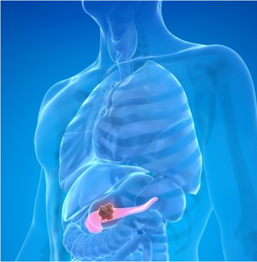
Translational Cancer ResearchCurrent chemotherapeutic agents are potent enough to kill cancer cells. Nonetheless, failure of chemotherapies for many cancers (e.g. breast and pancreatic cancers and various sarcomas) is primarily because these agents cannot reach cancer cells in amounts sufficient to cause complete cure. The abnormal microenvironment of these tumors drastically reduces perfusion and results in insufficient delivery of therapeutic agents. Tumor structural abnormalities is in large part an effect of mechanical stresses developed within the tumor due to unchecked cancer cell proliferation that strains the tumor microenvironment. Alleviation of these stresses has the potential to normalize the tumor, enhance delivery of drugs and improve treatment efficacy. We are testing the hypothesis that re-engineering the tumor microenvironment with stress-alleviating drugs has the potential to enhance chemotherapy. To explore this hypothesis, we make use of a mixture of cutting-edge computational and experimental techniques. We develop sophisticated models for the biomechanical response of tumors to analyze how stresses are generated and transmitted during tumor progression. We further perform animal studies to validate model predictions and identify the drugs that most effectively alleviate stress levels, normalize the tumor microenvironment and improve chemotherapy, nanomedicine and immunotherapy. Our research has revealed the mechanisms for stress generation and storage in tumors and led to new strategies for the use of anti-cancer therapies.
to mechanical stress. Protein extracts from MCS, mouse tumors and patient’s samples are subjected to proteomic analysis using Liquid Chromatography-Mass Spectrometry (LC-MS). Proteomic data are then analyzed to determine specific biological pathways and association analysis are employed to identify the most relevant mechanisms and markers of mechanoresistance. siRNAs and inhibitors against these markers are screened to find candidate drugs of mechanoresistance. |
In addition, in collaboration we pharmaceutical companies, we perform in vivo experiments in preclinical tumor models to test the efficacy of cancer therapeutics and the effectiveness of novel therapeutic strategies to improve cancer therapies. We combine principles and methods form bioengineering, medical imaging, pharmacology and tumor biology to advance cancer therapies from the bench to the bedside.
Biomarkers predictive of responseCancer therapies cause severe toxicities in some cases, underscoring the importance of biomarkers that can predict non-responders in a timely manner. To address this issue, we work under the hypothesis that certain biomechanical aspects of the tumour microenvironment, such as tumour stiffness, solid stress, perfusion and hypoxia, are responsible for the resistance to these therapies. Systemic administration of therapeutics requires a well-perfused vascular system, and that is often not the case in stiff and hypoxic tumours. Using clinically applied, ultrasound-based methods and computational biomechanical modelling in tumour-bearing mice and cancer patients, we aim to identify biomechanical biomarkers to predict therapy outcome. Specifically, apart from genomic/biological analysis, specific aspects of the mechanical microenvironment of tumors could be also employed as potential predictive biomarkers for immunotherapy.
|

|
SELECTED PUBLICATIONS
|