 |
OPTICAL DIAGNOSTICS LABORATORY |
“BERC epitomizes the multi-disciplinarity of cutting-edge biomedical research”Prof. Costas Pitris is a Professor at the KIOS Center of Excellence, Department of Electrical and Computer Engineering, University of Cyprus. He is heading the “Optical Diagnostics Laboratory” which he established in 2004. Prof. Pitris has completed his studies at the University of Texas at Austin (BS Honors in Electrical Engineering, 1993, MS in Electrical Engineering, 1995), Massachusetts Institute of Technology (Ph.D. in Electrical and Medical Engineering, 2000), and Harvard Medical School (MD Magna Cum Laude in Medicine, 2002). His main research interests include the areas of optical diagnostics, biomedical imaging and spectroscopy, as well as signal/image analysis, machine learning, and computational intelligence. Prof. Pitris has served as a PI or a co-PI in competitive research grants totaling over € 7 mil including a highly prestigious EU H2020 FET Open Grant, the first to be coordinated by a Cypriot institution. He is also one of the co-founders and a member of the executive committee of the KIOS Center of Excellence, which was the recipient of an EU H2020 TEMAING grant of over € 40 mil. Prof. Pitris has published 57 peer reviewed journal publications, 148 conference proceedings, 5 book chapters, and 1 book. He also holds 12 US, European and other patents. The citations to his work have reached more than 15600 (with an h-index of 41) according to Google Scholar. Professor Costas Pitris KIOS Center of Excellence Department of Electrical and Computer Engineering University of Cyprus cpitris-AT-ucy.ac.cy; (+357) 22 892297 |
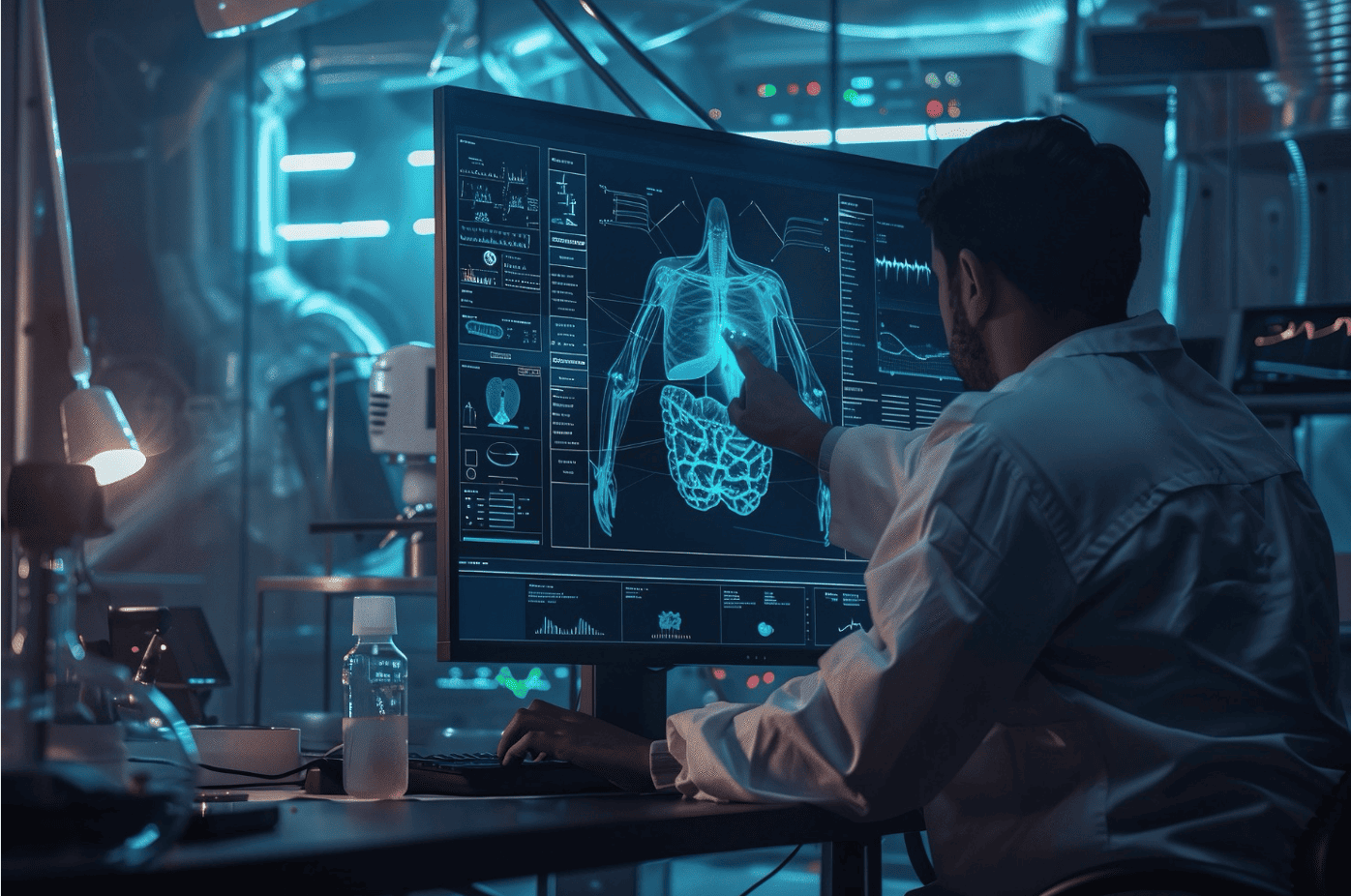
Enhancing the Capabilities of OCTOptical Coherence Tomography (OCT) is an emerging medical imaging modality which can provide microstructural images of tissue with a micrometer scale resolution. Despite this exquisite resolution, the images of the microstructure are not enough to identify the subtle changes associated with very early cancer. We aim to develop novel methods for the extraction of additional information from such images, improve the image contrast, and use Artificial Intelligence (AI) to accurately identify very early cancer using OCT images that provide the high accuracy required for population screening. Research in this area includes:
|
Artificial Intelligence-derived Biomarkers of DiseaseFor medical imaging to have a significant impact in the field of oncology, information must be extracted from the images and converted to biomarkers of disease that can be easily accessed and assessed by clinicians. These biomarkers should correlate with established and well-known biomarkers of disease and provide a reliable means of disease and risk diagnosis, stratification and even prediction. Image processing and machine learning are utilized to extract features that currently remain unseen and unused and reflect information regarding sub-resolution and biochemical changes in the tissue. These are being combined with other epidemiological information to create Information-Derived Biomarkers (IDBs) for early cancer detection. These IDBs are derived using both statistical inference but also informed machine learning (IML) to construct correlated and clinically meaningful biomarkers. Applications of state-of-the-art deep learning enhance the clinical utility of medical imaging but also provide explainable decision support. |
|
SELECTED PUBLICATIONS
|